Since 2011, UW students, faculty, and researchers have competed in the campuswide Cool Science Images contest and turned an artistic eye to their microscopes, scans, and schematics. With entries exploding with color and exciting patterns, this annual competition works to open people’s eyes to the beauty of science. And much of this UW research looks even better when you find out how it might advance medicine and improve life for people in Wisconsin and beyond. Read more about the science behind each winning image in the gallery below, then check out the 2023 winners.
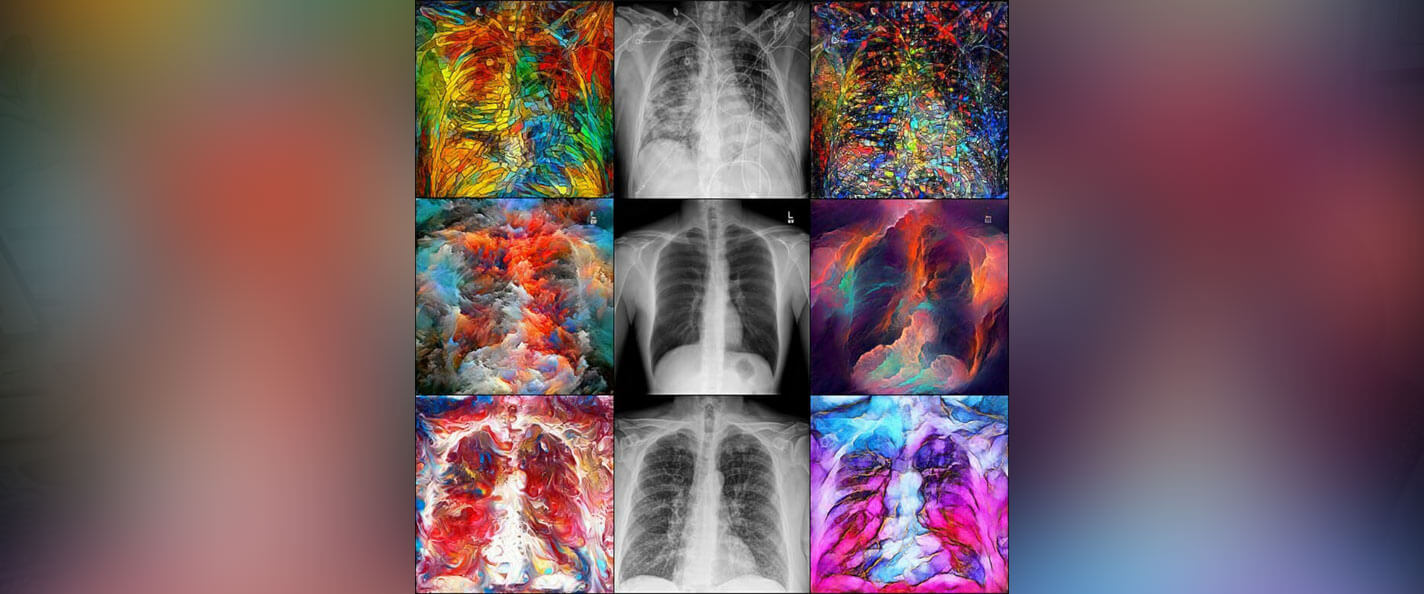
Generative adversarial networks, or GANs, are computing networks used in medical imaging. These grids compare X-rays of healthy lungs (center row) with those infected with COVID (top) and pneumonia (bottom). Image by 2022 contest winners Dalton Griner MS’19, PhD’23 and Xin Tie MS’21, PhDx’24.
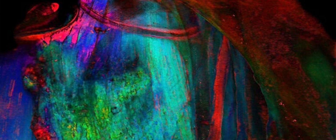
Did you know that a rat’s vagus nerve and neck muscles could look so pretty? A polarizing filter, demonstrated above, could help surgeons distinguish between types of fragile tissue when they operate. Image by 2022 contest winners Rex Chin-Hao Chen PhDx’24, Bruce Knudsen, Matthew LaLuzerne ’22, MS’23, MSx’24, James Trevathan, Kip Ludwig.
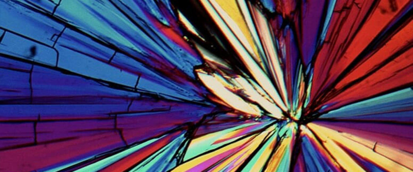
Arabitol, a sugar alcohol, takes on a stained-glass appearance with the use of a polarizer. The crystalline structure determines the effectiveness of a product, whether a pharmaceutical drug, LCD screen, or even chocolate. Image by 2022 contest winner Amy Neusaenger PhDx’24.
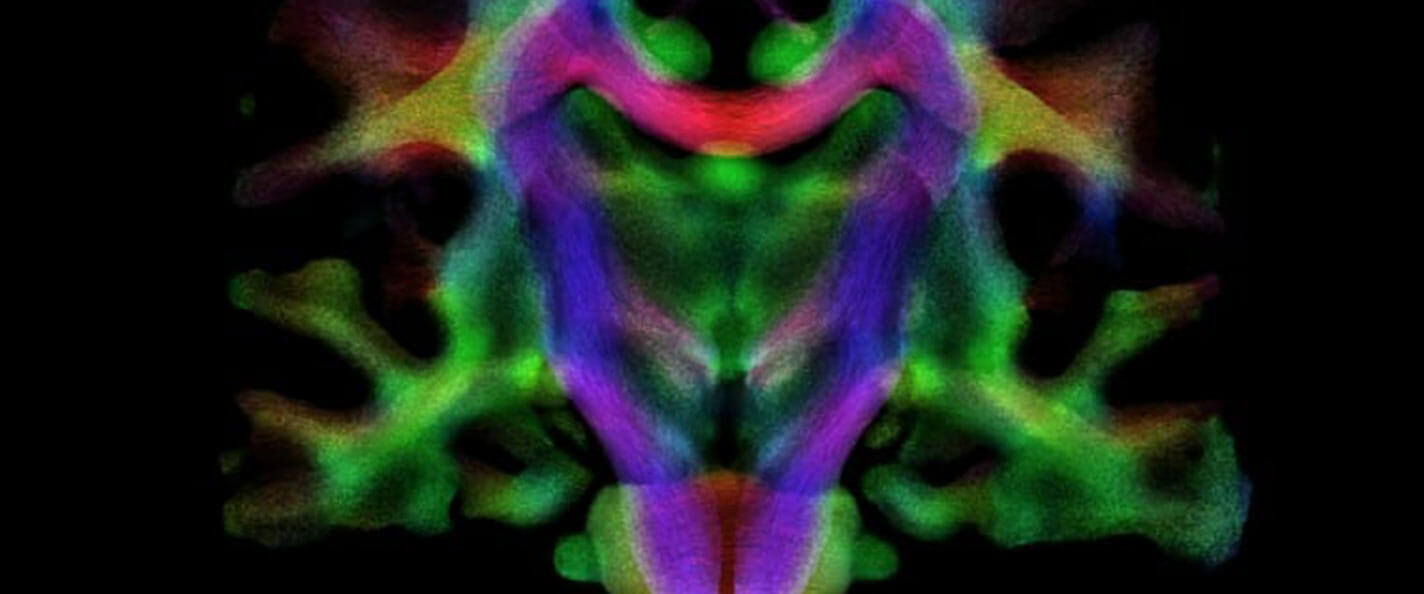
This kaleidoscopic view illustrates how white matter fibers, or connective tissue, are oriented throughout the human brain. Mapping out these fibers can help researchers understand brain development and neurological disorders. Image by 2021 contest winners Jose Guerrero MS’16, PhD’20 and Peter Ferrazzano.
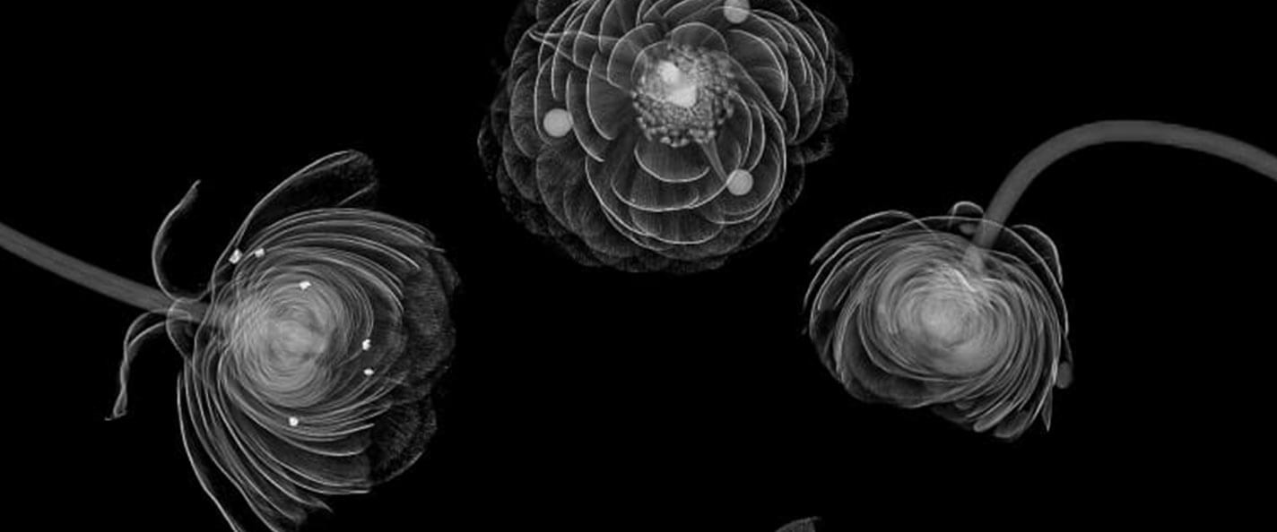
These flowers are dotted with tiny calcifications — much like breast tissue in the early stages of cancer. UW researchers are working to make it easier and faster to identify cancerous patterns, allowing patients to start treatment earlier. Image by 2020 contest winners Ran Zhang and Dalton Griner MS’19, PhD’23.
These human endothelial cells, embedded in a special hydrogel, are forming tunnels that may help insulin-producing islet cells survive longer after culturing. This gel could be key to a new therapy option for patients with diabetes. Image by 2019 contest winners Dan Tremmel ’10, PhD’22 and Vansh Jain ’19, MDx’24.
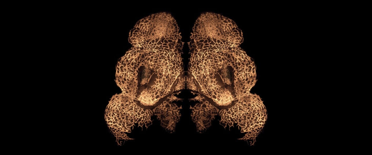
This micrograph shows blood vessels developing in the face of an 11-day-old mouse embryo. You’d see a similar face if you looked at a human embryo at 34 days. Image by 2018 contest winner Hannah Chung MS’18.
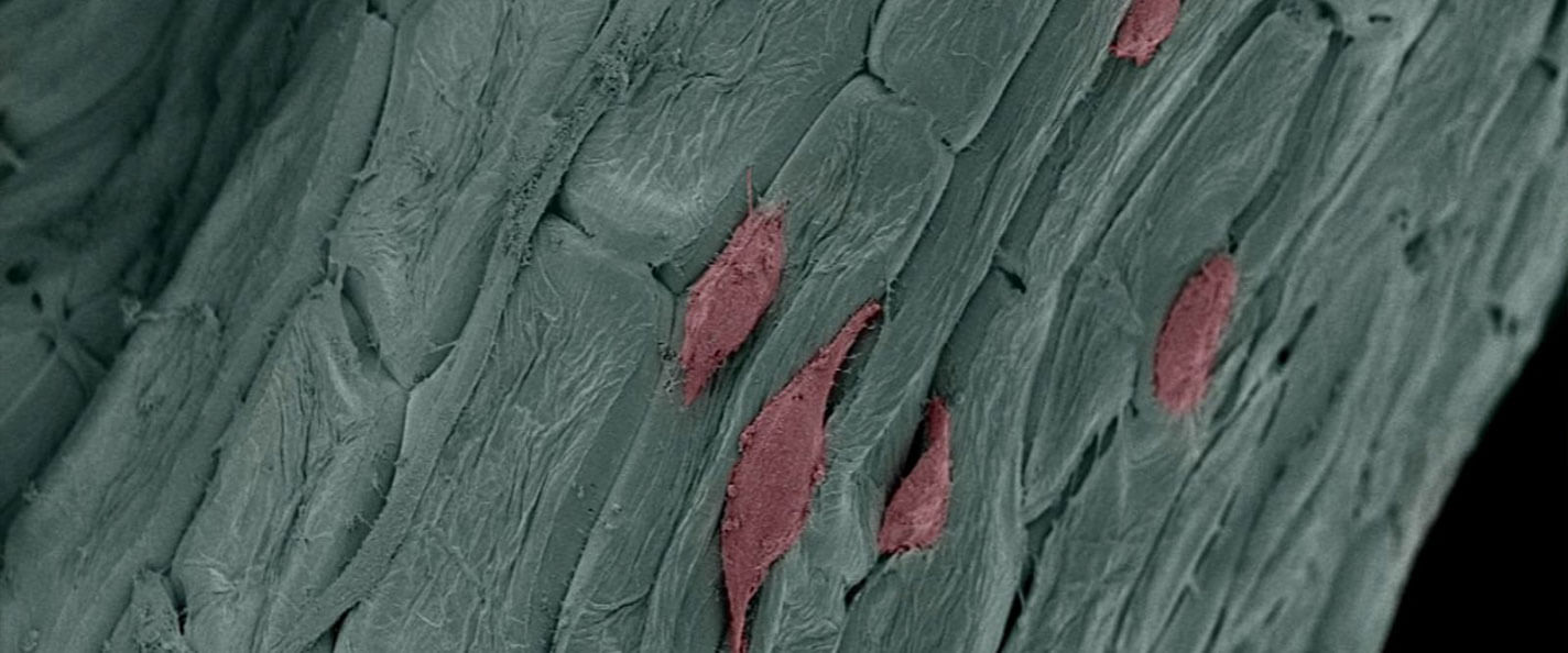
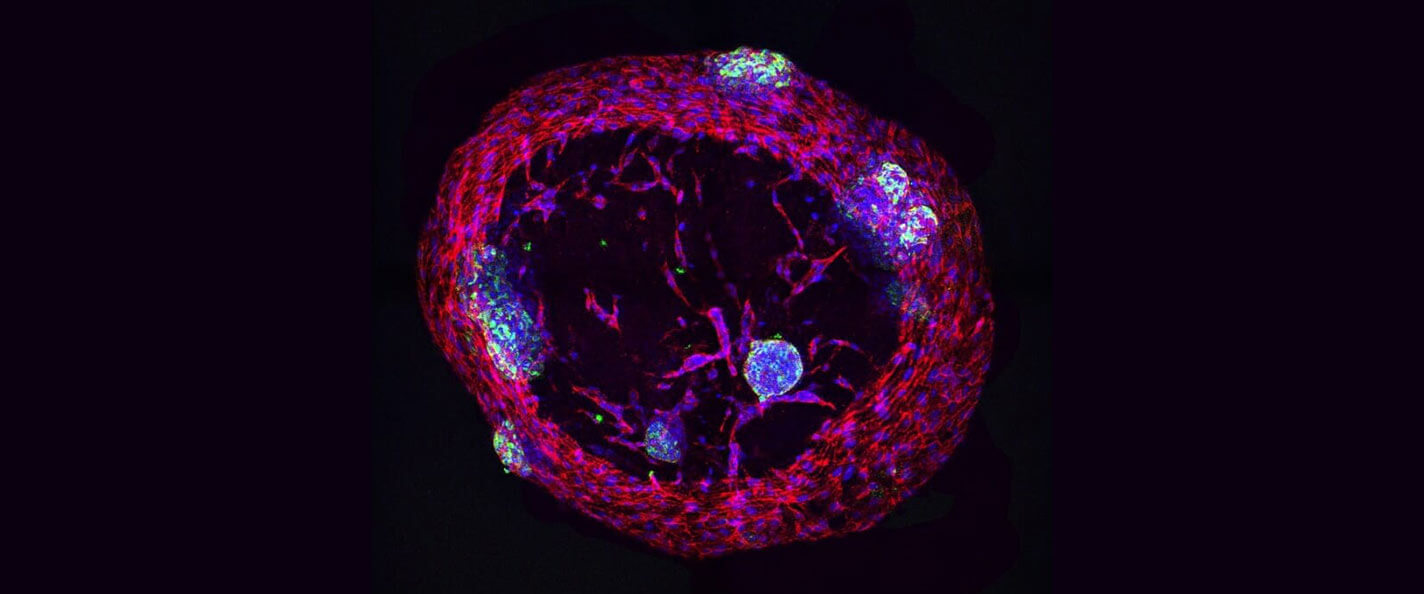
Parsley plus human fibroblast cells, shown in red, together form a base for growing stem cells. This scaffolding might one day work to repair bone and tissue. Image by 2017 contest winner Gianluca Fontana.
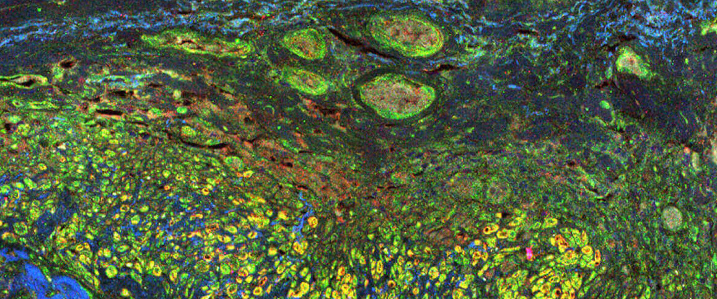
This Monet-like image resulted from staining antibodies and proteins that exist in human tissue. The technique helps researchers track protein activity throughout the immune response. By understanding how disease attacks the body, scientists can develop more effective ways to fight back. Image by 2016 contest winner Wei-Hua Lee.